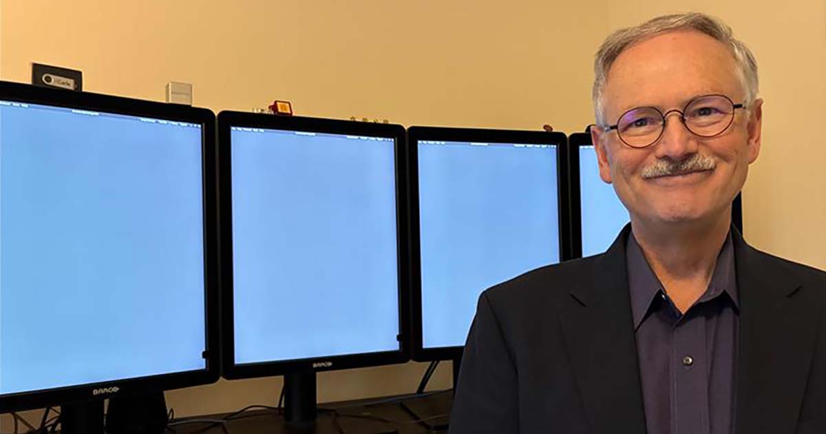
Dr. Doug W. Morton is a neuroradiologist at Carle Health and Carle Foundation Hospital, a Level 1 trauma center and comprehensive stroke center in Urbana, Illinois. He holds a PhD in neurobiology. A program lead for the Carle Enterprise Imaging Team, he directs a group in implementing and managing imaging informatics systems.
As an IT leader, physician and engineer, he believes medical technology – when designed with the end user in mind – can dramatically increase clinician engagement, quality care and workflow efficiency.
THE CHALLENGE
In 2018, Morton’s team started the search to replace Carle’s aging picture archiving and communication system. Carle had a “best of breed” approach to imaging technology, which meant the radiology PACS, transcription system, vendor-neutral archive and many other systems were from different vendors. At that time, Carle Health had just two hospitals – but five PACSs from different vendors.
“This technology model makes it harder for any clinician to have a comprehensive view of a patient’s imaging history,” Morton noted.
“For example, as a radiologist, I could not see a patient’s cardiology or ophthalmology images, a cardiologist could not see the patient’s radiology images, and so on. To create a more holistic view of a patient’s imaging record, we needed a consolidated repository – a VNA – for all imaging, including clinical photography, point-of-care ultrasound, scope images and digital pathology.
“The big question for us was: How big should we go with the PACS and VNA implementation?” he continued. “Our leaders advised me to solve the problem with something that could grow with our organization. I’m glad we took that approach, because in just five years, we expanded from two to eight hospitals and had to absorb and integrate nearly 900 terabytes of patient data, all sourced from disparate imaging systems and with no guarantee of data cleanliness.”
PROPOSAL
Carle Health needed a proven enterprise imaging system that could grow with the health system, and after careful evaluation, it selected Merge, which is composed of Merge VNA and Merge Universal Viewer, Morton recalled.
“This system could create a centralized repository of imaging data, readily accessed by users, regardless of the original source or format of the data,” he said. “This centralized repository would enable more efficient image sharing among clinicians and with the patients themselves.
“And, because Merge uses standard protocols such as DICOM and HL7, it could help enable seamless data exchange to support things like telemedicine, multidisciplinary team meetings and integrated care pathways,” he added.
The technology was pivotal to enabling Carle’s new enterprise imaging strategy of applying a more holistic approach across the growing organization. The vendor team also understood that in solving Carle’s initial problem, the health system also had new opportunities to optimize clinical workflows.
MEETING THE CHALLENGE
Carle Health used the enterprise imaging system to consolidate all imaging across the health system into one place. Clinicians now only need a single viewer to access a patient’s entire imaging history.
“One example of a specific clinical workflow we improved was giving full visibility to the patient’s history before they arrived for a cardiology exam,” Morton explained. “Now, because we have stored the data from the cardiology PACS into the VNA, we can use its pre-fetch algorithm to bring cardiology images into the cardiology PACS.
“The benefit of this is when patients show up for their cardiology appointment, those images are ready and available for the cardiologist without changing the existing cardiology workflow,” he continued.
The technology also helped staff integrate data that had some challenges. In some cases, data from incoming hospitals had fields filled in with wrong or unclear tags. Each hospital had different medical record numbering systems.
Data from outside sources, such as referring hospitals or emergency clinics, also was problematic. So, Carle staff created a “Holding Pen” – a temporary environment for receiving images from outside the system, where staff can review and properly tag images prior to patient admission, which is a valuable process for cleaning and integrating outside imaging studies.
“On the patient experience side, we integrated the universal viewer’s capabilities with our patient portal,” Morton noted. “Patients now can see their medical images alongside their doctors’ reports. We used an additional capability from another vendor to help explain medical terminology in everyday language so patients could better understand their reports. Usage of the portal has grown to about 11,000 patients each month who view their own images.
“Now that we have successfully integrated our PACS, cleaned up data labels and enabled patients’ access to their own images, we are working toward additional goals,” he continued. “For example, consolidating our ophthalmology data and clinical photography images. We’re also looking at developing a strategy for point-of-care ultrasound workflows and bringing digital pathology into a hybrid-cloud environment.”
RESULTS
When it comes to hard results, Morton points to four successes.
First, VNA images in the patient portal have been viewed by 11,000 patients each month.
“VNA image availability from the patient portal went live about one year ago,” he said. “Currently, about 11,000 individual patients view their images each month. Using this same functionality, patients can also download their pictures and send a link to out-of-network providers or institutions, allowing easy image sharing.”
Second, the VNA has been scaled to support system growth from three hospitals to eight hospitals.
“VNA scalability is critically important to support system growth,” Morton said. “The VNA technology provides a single scalable central image archive for our health system. When the VNA first went live, it supported three hospitals and about 200,000 studies annually. Our health system has now grown to eight hospitals and about 900,000 studies per year, with the VNA archive going from about 200 TB to over 900 TB.
“As our health system grew, the VNA was supplemented with additional resources, including storage and processing power,” he continued. “However, VNA function, responsiveness and architecture remained the same.”
Third, the VNA has provided a central archive without changing existing clinical workflows.
“The VNA prefetching assures patient data is delivered where and when needed,” he explained. “When a patient interacts with the healthcare system – for example, clinic appointment, ED visit, hospital admission, transfer and tumor conference – the appropriate image data is prefetched to the radiology and cardiology PACS.
“The central archive and prefetch strategy also allows clinical ‘load balancing,'” he added. “If a radiologist at one site is out of the office or overwhelmed, radiologists at other sites can pick up the load.”
This strategy also allows the team of night radiologists to use their existing workflow to read overnight cases from other hospitals in the system – cases previously sent to a nighthawk service. The night radiologist team currently reads about 7,000 cases per month that would have been sent to a nighthawk service because of the centralized archive provided by the Merge VNA.
And fourth, providers use a single image viewer for most clinical imaging.
“The enterprise viewer gives providers access to any image stored in the VNA via EHR links from each patient’s record,” Morton reported. “Before the VNA, most providers using a PACS viewer could access only radiology images. Now, all radiology and cardiology images from every hospital in our health system are stored in the VNA.
“We are in the process of rolling out clinical photography storage to the VNA,” he added. “The VNA photography app allows providers to use smartphones and tablets with the appropriate app to send photographs directly to the VNA linked to the correct patient with the proper anatomy and a study description. Our dermatology and scope imaging storage teams also simplify their photography needs using the VNA application.”
ADVICE FOR OTHERS
An enterprise approach to imaging has a lot of complex aspects – data integration, clinical workflows, security, clinician and patient trust, Morton said. It’s important to talk with the physicians in each department – radiology, cardiology, dermatology, ophthalmology and others – to get feedback on what they need from imaging systems to improve patient care, he advised. That’s the best way to get the needed information to optimize clinical workflows and meet their needs, he added.
“One other learning that is important to share is about the patient experience,” he said. “We wondered if having direct access to imaging would make patients more anxious about what certain images meant without the context a doctor can provide during a face-to-face encounter. But that hasn’t really happened.
“Patients just want to know their results and generally understand what the reports are telling them,” he concluded. “Rather than causing concern or anxiety, it has instead helped equip patients to have more meaningful conversations with their doctors.”
Follow Bill’s HIT coverage on LinkedIn: Bill Siwicki
Email him: bsiwicki@himss.org
Healthcare IT News is a HIMSS Media publication.
WATCH NOW: Chief AI Officers require a deep understanding of the technologies and clinical ops
